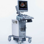
Following your ablation procedure, you will receive follow-up evaluations. Patients are followed at approximately one week and three months. We also like to evaluate at six months and one year post-procedure. These evaluations usually require a twenty minute visit to rescan the treated veins using ultrasound. By this time, patients are familiar with ultrasound imaging. The initial appointment with Minnesota Vein Center, Dr. Pal began with a review of the patient’s medical history and review of symptoms the patient had been experiencing. Next, a focused physical exam was performed to thoroughly check for abnormalities present in the veins. Dr. Pal was evaluating the patient for the following:
- Visible tiny blue or red veins
- Bulging veins (above the skin surface)
- Skin discoloration or skin thickening
- Warmth
- Open or closed leg wounds
- Ankle or leg swelling
Once the physical exam was completed, ultrasound imaging, a simple, noninvasive procedure, was used to get a more in-depth look at vessels. Using high frequency sound waves, pictures were created to show the structure of the suspected diseased veins. Movement of blood was also seen during ultrasound within these veins. Using ultrasound, it was easy to identify any narrowing of the venous walls or functional problems of the valves within the veins. Ultrasound imaging is especially important for those patients with venous disease who have had no obvious physical signs. At your post-procedure follow-up ultrasound evaluations, you will feel more familiar with the ultrasound exam process. You may even be interested to observe and to discover the outcome of your successfully treated ‘problem vein.’ At one week, we are able to determine that the vein remains closed. At three months, again the closed vein is verified. After six months, we most often see the vein has been reabsorbed by the body. As venous disease IS a disease process, follow-up evaluations allows the vein specialist, Dr. Pal a time to examine by ultrasound, the treated vessels and to identify newly visualized problematic veins. Follow-up and early detection provide opportunity to prevent further venous disease, a condition that is heredity-related and known to recur in other vessels.

